
See More In Less Time: Line REscan AION and Kinetix
Ando Zehrer and Dušan Popov-Čeleketić
Teledyne Photometrics and Confocal NL
The combination of Line REscan AION confocal system and Kinetix camera offers unparalleled capabilities for live cell imaging. AION with its motorized switchable pinhole, provides spot-on confocality for a wide range of objectives with a large field of view. Kinetix delivers unmatched sensitivity and speed with a quantum efficiency (QE) higher than 95%. This combination is ideal for live cell imaging as it enables high-speed image acquisition of large samples, enhanced by real-time deconvolution and reduced phototoxicity. This adaptable and user friendly system supports simultaneous dual-camera imaging and seamlessly integrates with microscope bodies and software, offering an unprecedented performance in confocal microscopy.
Introduction
Modern microscopy is a vital tool for pushing the boundaries of human knowledge in the life sciences, advancing one step further with every new development in hardware or software. Spinning disk confocal microscopy is a popular imaging method within the life sciences due to the optical sectioning providing improved resolution and sharper images within single focal planes. However, current live cell spinning disk confocal microscopy faces limitations, creating a demand for more gentle imaging with higher resolution, improved acquisition rates, increased spectral sensitivity, a larger field of view (FOV) and more.
A disruptive concept has been put forward with the Line REscan AION confocal unit from Confocal NL, enabling high resolution imaging in a compact and cost-effective solution. A crucial part of any imaging setup is the detector, and the Kinetix from Teledyne Photometrics is a groundbreaking modern back-illuminated sCMOS camera with a balance between speed and sensitivity, while providing one of the largest commercially available sensors for large FOV imaging.
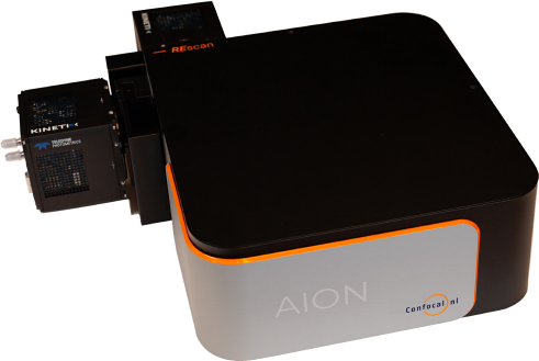
Figure 1: Line REscan AION confocal microscope equipped with two Kinetix sCMOS cameras for multicolor imaging, with a speed of over 100 fps across a 3k x 3k pixel array.
In this article we aim to highlight the powerful integration of these two technologies, emphasizing the unique advantages they provide to microscopy. The result is an engineered solution enabling high-speed imaging on up to two detectors simultaneously, across a large FOV, all with the resolution and axial sectioning of a state-of- the-art confocal system.
Limitations Surpassed by the New Setup
Typical sCMOS cameras often lack the sensitivity and the acquisition speeds necessary for applications where weak signals must be detected at high speeds. Using the Kinetix results in over double the acquisition speed, and the high QE of >95% ranging from visible to near-infrared (NIR) wavelengths with minimal levels of noise (< 1 e- read noise) alleviates bottlenecks encountered with previous detectors. The Kinetix sensor (3200 x 3200 pixels, 6.5 µm pixel size) provides flexibility when choosing magnification settings as well as other imaging modalities. The physical size of the chip (29.42 mm diagonal, i.e. FN29) captures far more data in every frame than typical sCMOS cameras, but requires a suitably large FOV system to pair with in order to maximize the benefits of the large sensor.
The AION has been optimized to image large samples and fully leverages the large FOV and the resolution provided by the Kinetix, thus providing over double the FOV size of conventional systems. This instates the hardware combination of AION and Kinetix as the method of choice for a plethora of different microscopy applications, taking advantage of the larger FOV or the accompanying increased throughput provided by high speed imaging.
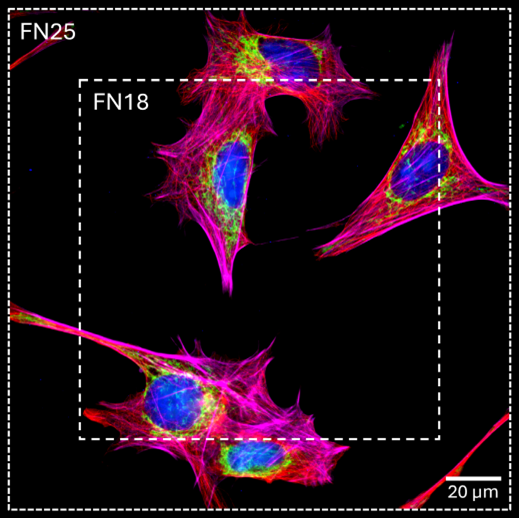
Figure 2: Comparing a FN18 system with the FN25 provided by the AION and Kinetix, using a 100x 1.50 NA objective. Image shows fixed human fibroblast cells, nucleus - DAPI (blue), mitochondria - Tom20-Alexa488 (green), microtubules - Tubulin- Alexa555 (red), actin – Alexa647 (magenta).
AION is designed to be fully diffraction limited at the FOV with a diagonal of 25 mm, FN25, for objective apertures corresponding to 60x 1.4 NA and smaller. That means that the value NA/Mag of an objective must be smaller or equal to 1.4/60 to provide the diffraction-limited image using FN25 FOV. Thus, using high magnification and high NA objectives (such as 100x, 1.45 NA) will enable imaging double the sample surface when imaged on a system enabling FN25 FOV, when compared to more common FN18 systems (Fig.2). AION is also equipped with a switchable pinhole, achieving optimal confocality for a wide range of objectives (4x-100x).
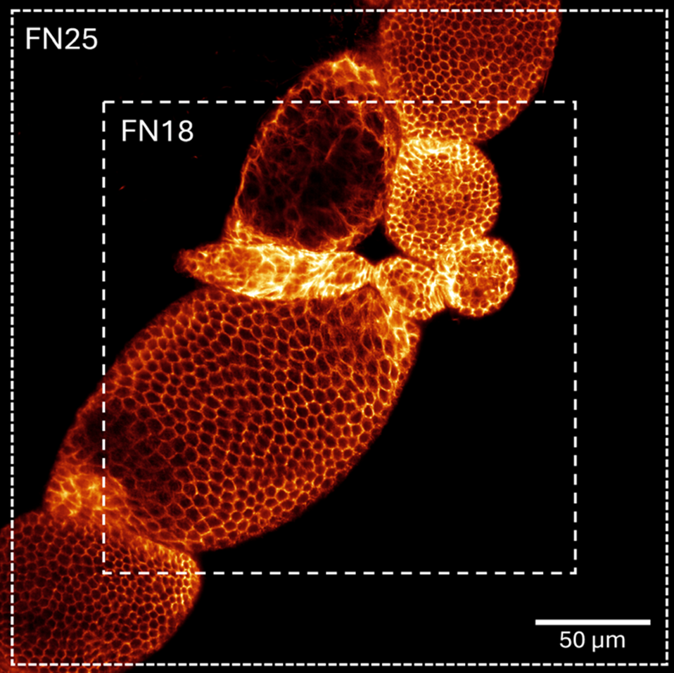
Figure 3: Comparing a FN18 system with the FN25 provided by the AION and Kinetix, using a 60x 1.42 NA objective. Image shows D. melanogaster oocytes, microtubules – Alexa 555.
Compared to its predecessors, the optimized optics and the smart imaging capabilities of AION, combined with the speed and sensitivity of the Kinetix improves the quality and throughput of confocal microscopy. The AION has three different versions (α, λ and Σ) differing in hardware configuration, which can all be adapted to every common microscope output. The design even accounts for simultaneous imaging of different channels by incorporating two cameras with a large FOV. When used with two Kinetix cameras, over 100 frames per second of a 25 mm diagonal FOV (450 x 450 µm2 with 40x objective) can be imaged per channel. A real-time resolution of 170 nm can be achieved using deconvolution on the fly, producing high-quality images at depths beyond 500 µm. The motorization of the pinhole further increases imaging quality, with other motorization and automation features promoting improved user-friendliness.
This flexibility is further supported by the illumination provided with the AION which can have up to twice the number of excitation lines, even ranging into the NIR (400-1100 nm). The high imaging depth is also a big advantage of the system, which can be enhanced by the usage of longer wavelengths for reduced aberrations or scattering in deeper samples. Despite being a confocal system, the illumination pattern has been improved to have an several-fold reduced phototoxicity compared to conventional systems. This feature is highly relevant for live-cell applications which could be conducted for longer durations and be more reliable due to the reduced damage cells sustain during an experiment. The installation of the setup is user-friendly as it implements all elements with open source software such as Micro-Manager. Drivers are available to incorporate the hardware in other control software or to create a custom solution.
Conclusion
The new Line REscan AION system reaches the pinnacle of confocal technology in combination with up to two Kinetix cameras (Fig. 1). The simultaneous acquisition of such a large FOV combined with high temporal and spatial resolution, real-time deconvolution, and high imaging depth, provides an imaging tool with unprecedented capabilities. The system is very adaptable thanks to its simple hardware and software implementation to available microscopes and its compact build. The AION-Kinetix combination is unmatched as a cutting-edge high-quality confocal system.

Acknowledgments
Images were taken using AION Line REscan confocal microscope in combination with a Kinetix camera. The authors are grateful to Janine Cravo, Anna Fehér, Ahmad Farhan Bhatti and Spyridon Theodoros Goudelis for their help with the generation of figures.