
Magneto-Optical Kerr Effect (MOKE) Imaging
Prof. Alejandro Silhanek, Dr. Emile Fourneau
Quantum Materials, Department of Physics, University of Liège, Belgium
Background
Prof. Alejandro Silhanek leads the Experimental Physics of Nanostructured Materials research group, which focuses on the study of magnetic, superconducting, and hybrid materials. This involves both fabrication via electron beam lithography and material characterization through magneto-optical imaging, specifically magnetic domains imaging using the magneto-optical Kerr effect (MOKE).
Post-doctoral researcher Dr. Emile Fourneau described his work, “When you are working at small dimensions, the way the magnetic domains arrange is essential for understanding the macroscopic behaviour of the material and has consequences for its potential technological applications. To understand this, you need to visualize magnetic domains and their arrangement. However, at this microscopic scale, the optical signal can be very low, and one needs the state-of-the-art instrumentation to be able to pick up such tiny signals.”
“We are using magneto‑optical Kerr microscopy to visualise the magnetic domains in micrometer or nanometer structures. Besides magnetic materials, we are also working with superconductors and using the Faraday effect to see the magnetic field penetration through the material as it penetrates and exits from the superconductor.”

Figure 1: A comparison of optical (tomography) imaging (left) and magneto-optical Kerr effect (MOKE) imaging (right). Each square is 25 µm in size, and images were obtained using a 50x microscope objective with the Prime BSI Express sCMOS camera.
Challenge
Dr. Fourneau explained the imaging challenges he faces in this line of research, “We are imaging through a series of polarizers/analyzers in cross configuration, so the intensity of the light reaching the camera is very low, and the contrast induced by the Kerr effect is about 0.1% or 0.01% of the detecting light intensity. To be able to obtain a high-quality image, one must use a camera that is very sensitive and has low noise. Our previous CCD camera had too much noise, making the image blurred and grainy.”
Another challenge is that our setup is mounted on a cryostat, which introduces more noise due to additional windows that depolarize the light and vibrations caused by cryopumping. Additionally, our goal is to image few-atomic-layer-thick films at very low temperatures, which means the signal we receive is nearly non-existent. In order to overcome these challenges, we need a camera with a very high signal-to-noise ratio.
In this application, sensitivity is paramount, requiring a camera with both high signal collection capability and low noise levels.
Solution
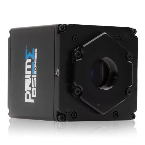
The Prime BSI Express sCMOS camera offers the ideal solution for this application, featuring near-perfect 95% quantum efficiency and very low read noise.
Dr. Fourneau told us about his experience with the camera, “With the new CMOS Prime BSI Express camera, it’s about 10 times better visually than our previous CCD camera in terms of Kerr effect contrast; now we can extract the signal and obtain a high-quality image, which is key for scientifically proving our claims. We were not seeing any signal before, and now with the Prime BSI we are close to publishing our work.”
“Every spec is better: the resolution, bit depth, and acquisition time. With this camera, we can obtain a good signal 100 times faster. What’s also very interesting is the capability to control the camera using a Python script. It was easy to set the camera up, and the customer support documentation was clear; we achieved exactly what we wanted very rapidly.”
Learn More About The Prime BSI Express