Live Nematode Imaging Shows Intergenerational Link to Diet
Sigma Pradhan, Klement Stojanovski, Ferdinand Dellemann, Sacha Psalmon, Joel Tuomaala, Nicholas E. Stroustrup, Benjamin D. Towbin
Institute of Cell Biology, University of Bern, Bern, Switzerland
Background
Every cell must balance its protein production capacity against its nutritional environment. Ribosomes (responsible for synthesising proteins within the cell) are among the most metabolically costly cellular components to produce, and their abundance scales closely with growth rate across organisms from bacteria to mammals. When nutrients are scarce and growth is slow, cells downregulate ribosome production to avoid wasteful expenditure. In the nematode worm Caenorhabditis elegans, dietary restriction is well established to reduce ribosome expression and slow organismal growth within a generation.
However, a deeper and largely unexplored question concerns what happens across generations: when a mother experiences dietary restriction, is the proteome of her offspring simply reset at the moment of fertilisation, or do maternal nutritional conditions leave a lasting imprint on the molecular composition of the next generation?
Researchers at the University of Bern addressed this question by combining tandem-mass-tag (TMT) proteomics with high-resolution live fluorescence imaging in C. elegans, tracking ribosomal protein levels within and across generations, and dissecting the cellular and signalling mechanisms by which maternal physiology influences offspring growth capacity. A central goal was to determine whether the maternal germline acts as the conduit through which nutritional information is transmitted to progeny.
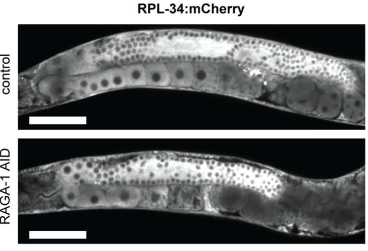
Figure 1: Representative image of RPL-34:mCherry fluorescence in control and auxin treated animals. Obtained with the Kinetix sCMOS, scale bar = 100μm.
Challenge
The team used a dual-camera setup with a spinning disk confocal microscope for live-cell fluorescence imaging. This research involved imaging live C. elegans with fluorescently tagged ribosomal proteins (RPL-34:mCherry and RAGA-1:GFP:AID), imaging directly within the maternal germline. The germline is a highly dynamic 3D tissue in which individual oocytes at different stages of maturation must be resolved and quantified with precision. The C. elegans germline is organised along a spatial gradient and the University of Bern team needed to detect subtle but biologically meaningful differences in fluorescence intensity across specific regions of this structure to determine where and how ribosomal protein levels respond to mTORC1 perturbation.
Biological differences were modest, only ~10% change in fluorescence intensity, requiring an imaging system with exceptional sensitivity, quantitivity and reproducability.
This demanded a camera capable of delivering high sensitivity and low noise at the single-cell level, operating on a spinning disk confocal platform to achieve the optical sectioning necessary to image deep within the worm's gonad. The combination of inherently low signal from endogenously tagged proteins expressed at physiological levels, the need for spatial precision across oocytes of varying size, and the requirement to detect graded changes in fluorescence intensity across the proximal-to-distal germline axis made detector performance a critical determinant of data quality.
Solution
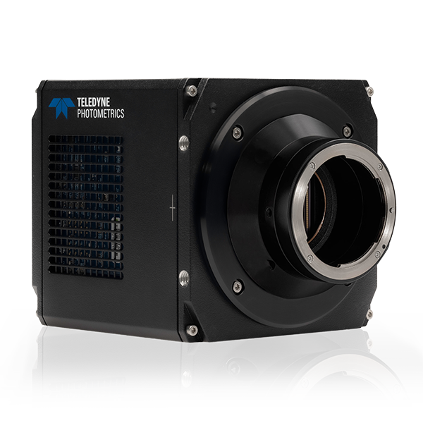
The dual-camera system uses two Kinetix sCMOS cameras on a Yokogawa W1 large field spinning disk system, a powerful solution and ideal for sensitive fluorescence imaging of live samples. Kinetix provides low read noise and high collection efficiency for imaging, combined with high readout speed for synchronising to a spinning disk. This enabled the Bern team to image fluorescently labeled ribosomal proteins within the germline at high spatial resolution, with accurate quantification of small changes in fluorescence intensity to distinguish biologically meaningful differences in protein abundance.
Kinetix also supported automation of large dataset acquisition and analysis, with consistent fluorescence images enabling computational segmentation and quantitative analysis of individual oocytes, embryos, and germline regions, allowing researchers to correlate maternal physiology with protein provisioning in offspring.
Along with spinning disk imaging, Kinetix was also used for widefield time-lapse imaging, revealing that maternal dietary restriction reduces ribosomal proteins offered to offspring, slowing early larval growth before normal ribosome levels are restored. These findings demonstrate how sensitive quantitative imaging can uncover subtle developmental mechanisms that would be difficult to detect using conventional imaging approaches.
By delivering high sensitivity, excellent quantitative performance, and reliable image quality, the Kinetix camera enabled researchers to visualize the molecular basis of intergenerational inheritance and generate robust datasets supporting this important biological discovery.
Reference
Sigma Pradhan, Klement Stojanovski, Ferdinand Dellemann, Sacha Psalmon, Joel Tuomaala, and Nicholas E. Stroustrup (2026) Dietary restriction shapes intergenerational ribosome abundance and early growth of Caenorhabditis elegans offspring. Plos Biology. 2026 Apr;24(4):e3003692. DOI: 10.1371/journal.pbio.3003692. PMID: 41955178; PMCID: PMC13065021.