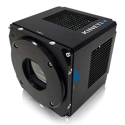
Kinetix22 and the THUNDER Imager Cell
Investigating 2D Cultures, Organoids, and Whole Organisms using the Kinetix22 sCMOS camera with a THUNDER Imager Cell system
Summary
Widefield fluorescence microscopy remains an attractive approach for live‑cell and large‑area imaging due to its speed, gentle illumination, and experimental simplicity. However, imaging optically thick or highly scattering samples - including dense 2D cultures, 3D organoids, and whole organisms - is fundamentally limited by out‑of‑focus background, reduced contrast, and phototoxicity when higher illumination intensities are required.
This application note presents a widefield imaging workflow that addresses these challenges by combining the Leica Microsystems THUNDER Imager Cell system with the Teledyne Photometrics Kinetix22 sCMOS camera, which delivers 95% quantum efficiency (QE) and sub‑electron read noise. By pairing high‑sensitivity, large‑field detection with THUNDER computational clearing to remove out of focus blur, this approach enables high‑contrast, high‑speed 2D and 3D imaging of challenging samples at low light doses. Performance is demonstrated across fixed and live biological samples, ranging from cultured cells to neural organoids and live zebrafish embryos.
Introduction
Live‑cell imaging of large and structurally complex biological samples places competing demands on conditions for microscopy experiments. Such studies often require large fields of view, sub‑cellular resolution, high‑sensitivity, and fast acquisition, all while maintaining low illumination doses to enable reliable quantification over extended time periods. For these reasons, widefield microscopy is an attractive approach.
However, optically thick and heterogeneous samples – such as dense cell cultures, organoids, and living organisms – generate substantial out‑of‑focus fluorescence in widefield imaging, reducing contrast and quantitative accuracy. While optical sectioning techniques can mitigate these effects, they can introduce tradeoffs in speed, phototoxicity, or experimental complexity. Widefield microscopy therefore remains highly beneficial, provided that background fluorescence can be effectively suppressed.
Optimizing imaging parameters for live-cell imaging
> Sensitivity depends on maximizing sample signal intensity and camera signal collection, while minimizing noise such as sample background and camera read noise. Although signal levels can be increased by using more intense illumination or longer exposure times, these approaches may lead to photobleaching and phototoxicity, compromising sample viability and limiting imaging speed. The challenges associated with low‑signal samples can be overcome by the combination of THUNDER computational clearing and the high quantum efficiency (QE) and low read noise of the Kinetix22.
> Spatial resolution is determined by both microscope optics and camera pixel size. The optical component is addressed through the compatibility of THUNDER Imager Cell with a wide range of Leica microscope objectives, together with Smart Correction Collar (SmartCORR) technology, which provides hands‑free optimization of objective correction collar settings to match the experiment. This improves image quality by reducing spherical aberrations. Cameral pixel size is addressed by the Kinetix22’s 6.5 µm pixels, a balanced pixel size optimized for use with both high‑ and low‑ magnification objectives, enabling sub cellular resolutions even across large samples at low magnification.
> Imaging speed is largely determined by the camera and is influenced mostly by the exposure time and the digital interface with the PC. The Kinetix22 camera can operate at over 660 frames per second (fps) in Speed mode, and over 110 fps in modes optimized for Sensitivity and Dynamic Range. Combined with its high sensitivity and the latest PCIe (Peripheral Component Interconnect Express) cable, the Kinetix22 enables acquisition of high‑contrast images even at short exposure times, supporting truly high‑speed imaging.
> Field of view (FOV) depends on the combined performance of the camera and microscope system. The Kinetix22 and THUNDER Imager Cell feature a matching 22 mm FOV which enables the capture of more data in every frame compared to configurations with mismatched optical fields. The Kinetix22 is one of the only cameras that can fully utilize the FOV of the THUNDER Imager Cell.
Combining THUNDER imager Cell with the Kinetix22
The THUNDER Imager Cell and Teledyne Photometrics’ Kinetix22 camera provide a high speed, high performance imaging platform. The high sensitivity of the system supports low light imaging, helping to minimize phototoxicity and preserve physiologically relevant data. The Kinetix22’s 22 mm field of view, 95% quantum efficiency, and sub electron read noise enable efficient photon collection across large samples.
While careful optimization of sensitivity, resolution, speed, and FOV is essential, these parameters alone cannot fully overcome the fundamental optical limitations imposed by thick and scattering samples when using widefield imaging. A key remaining challenge is background haze from out‑of‑focus light. In thick or scattering samples such as organoids or tissues, this background noise reduces contrast, signal‑to‑noise ratio (SNR), and quantitative accuracy. This is especially problematic in live‑cell experiments where low light doses are required.
This is where Leica Microsystems THUNDER technology plays an important role, enhancing image contrast in real time whilst still enabling users to benefit from the speed and gentle illumination conditions typical of widefield systems.
THUNDER Computational Clearing
At the heart of THUNDER is Computational Clearing (CC), Leica Microsystems patented background subtraction method that separates in‑focus signal from out‑of‑focus blur in real time. Unlike traditional deconvolution methods that redistribute all light detected in an image (including scattered light), Computational Clearing efficiently differentiates between signal and background. By excluding unwanted background, this approach delivers more realistic representations of 3D structures and generates high resolution, high contrast images.
THUNDER computational clearing can either be applied on‑the‑fly as part of the acquisition, or in post‑processing. THUNDER offers three clearing modes:
> Instant Computational Clearing (ICC): Enables single‑image haze removal for live or dynamic samples.
> Small Volume Computational Clearing (SVCC): Adds 3D deconvolution tailored to thin specimens.
> Large Volume Computational Clearing (LVCC): Adds 3D deconvolution optimized for thick samples such as organoids and tissue sections, to improve contrast deeper into large volumes.
THUNDER preserves quantitative fluorescence intensity relationships, maintaining a linear response across exposure times and signal intensity levels. It delivers consistent performance across samples with heterogeneous background fluorescence levels. By enabling clearer visualization deeper within large‑volume samples than conventional widefield methods, THUNDER reveals structural details that would otherwise be obscured by out‑of‑focus light, thereby making thick samples accessible for robust analysis.
Here, the performance of the THUNDER Imager Cell with Kinetix22 sCMOS camera is demonstrated across a range of large 2D and 3D biological applications, illustrating the impact of THUNDER on diverse sample types.
Results
2D Imaging of U2OS Cell Cultures: Large FOV Imaging
In the first example, a THUNDER Imager Cell with Kinetix 22 sCMOS camera was used to image an entire U2OS cell population across a standard microscope slide at either 10x (Figure 1) or 20x (Figure 2) magnification. Crucially, high spatial resolution was maintained, even with the low 10× magnification, as shown in the magnified inset images in both Figures 1 and 2. This provided sub‑cellular resolution across the full field, enabling imaging of very large cell populations while preserving fine structural details.
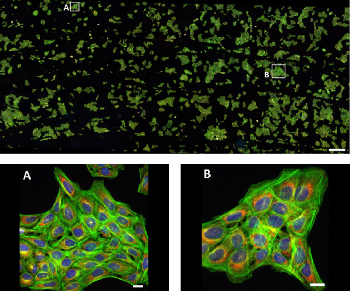
Figure 1: Large field of view (FOV) image of U2OS human osteosarcoma cells stained with DAPI (nuclei, blue), Alexa Fluor 488 Phalloidin (F‑actin, green), MitoTracker Red (mitochondria, red), and Alexa Fluor 647 Anti‑tubulin (tubulin, white). Acquired on a THUNDER Imager Cell with 10x magnification using automated XY tiling and stitching. Magnified insets are indicated by white squares on the main image, corresponding to A and B. Main image scale bar 100 µm, inset scale bars 10 µm. U2OS cells provided by Dr. Niloufer Irani (Micron Bioimaging Facility, University of Oxford).
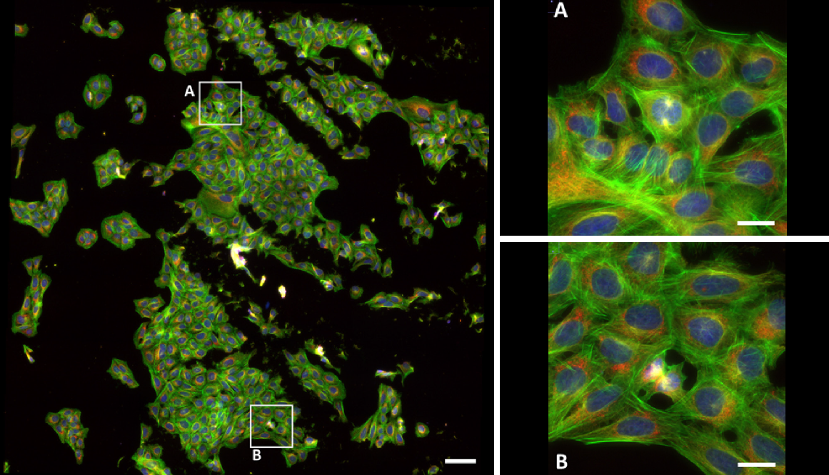
Figure 2: Large field of view (FOV) image of U2OS human osteosarcoma cells stained with DAPI (nuclei, blue), Alexa Fluor 488 Phalloidin (F‑actin, green), MitoTracker Red (mitochondria, red), and Alexa Fluor 647 Anti‑tubulin (tubulin, white). Acquired on a THUNDER Imager Cell with 20x magnification using automated XY tiling and stitching. Magnified inserts indicated by white squares on the main image, corresponding to A and B. Main image scale bar 50 µm, insert scale bars 10 µm. U2OS cells provided by Dr. Niloufer Irani (Micron Bioimaging Facility, University of Oxford).
2D Imaging of U2OS Cell Cultures: Sub-cellular Resolution Imaging
With low magnifications, such as the 10x and 20x shown previously in Figs 1 and 2, the THUNDER Imager Cell and Kinetix22 can maximize FOV with slightly reduced spatial resolution as the objectives have a lower numerical aperture (NA). When switching to higher magnifications, such as 40x or 63x water immersion, spatial resolution can be maximized thanks to Nyquist sampling with the Kinetix22’s 6.5 µm pixel size. This is shown in Figure 3.
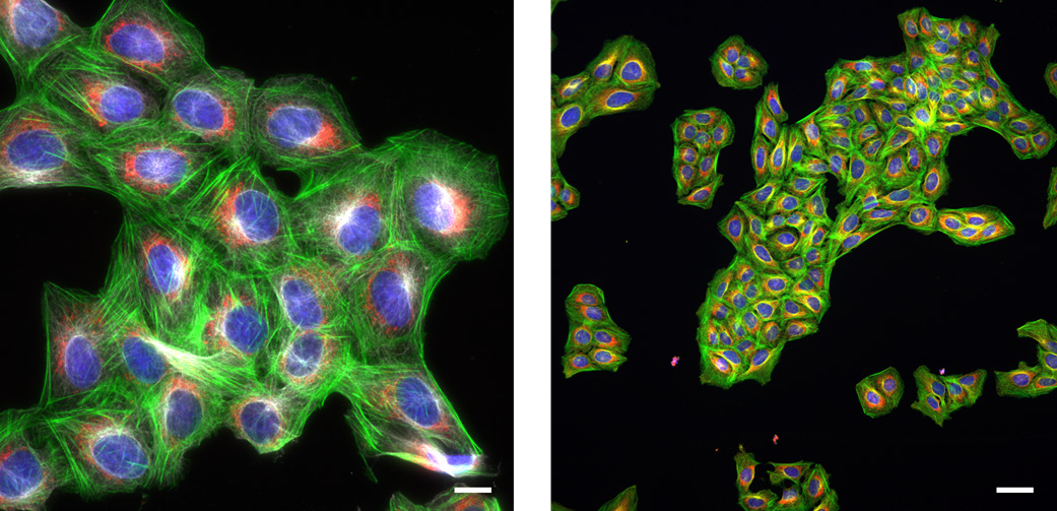
Figure 3: High resolution, high magnification images of U2OS human osteosarcoma cells, stained with DAPI (nuclei, blue), Alexa Fluor 488 Phalloidin (F‑actin, green), MitoTracker Red (mitochondria, red), and Alexa Fluor 647 Anti‑tubulin (tubulin, white). Acquired on THUNDER Imager Cell with 63x water immersion magnification (left, scale bar 10 µm) and 40x magnification (right, scale bar 50 µm). U2OS cells provided by Dr. Niloufer Irani (Micron Bioimaging Facility, University of Oxford).
2D Imaging of U2OS Cell Cultures: Contrast Improvements with THUNDER
With THUNDER Imager Cell, a further increase in contrast and feature identification is made possible using THUNDER computational clearing technology. This is demonstrated in Figure 4 where the original widefield image is compared to the THUNDER computationally‑cleared image. Both images also feature a line profile in the same location, demonstrating the changes in quantitative signal values and the signal-to‑background noise levels following the THUNDER clearing process.
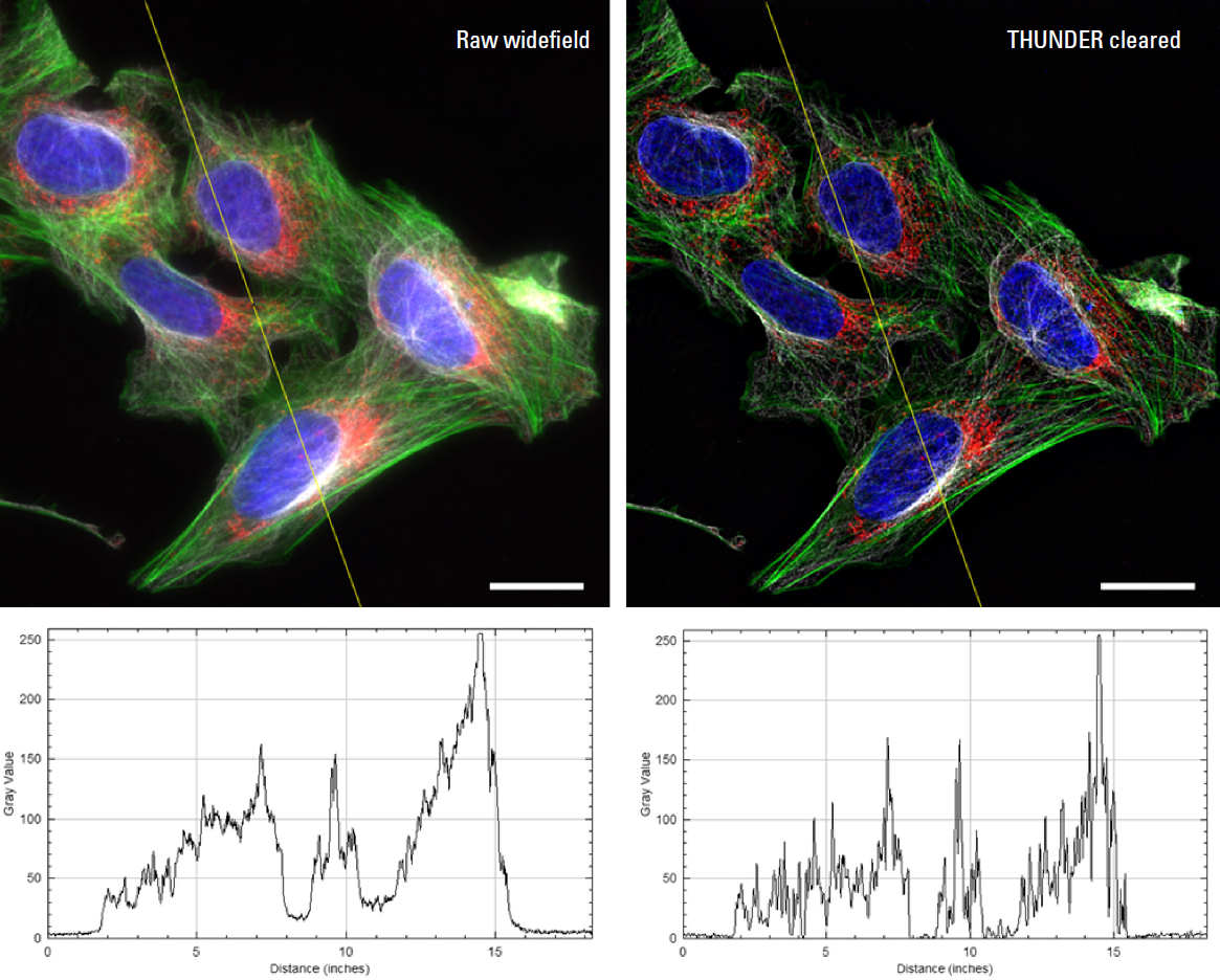
Figure 4: High resolution, high magnification images of U2OS human osteosarcoma cells, stained with DAPI (nuclei, blue), Alexa Fluor 488 Phalloidin (F‑actin, green), MitoTracker Red (mitochondria, red), and Alexa Fluor 647 Anti‑tubulin (tubulin, white). Acquired on THUNDER Imager Cell with 63x water immersion magnification. Left: Raw widefield image, Right: THUNDER cleared image. Scale bars are 20 µm. Below are line profiles across the yellow line through each image in the same location. U2OS cells provided by Dr. Niloufer Irani (Micron Bioimaging Facility, University of Oxford).
The improvements in image quality and resolution delivered by THUNDER enabled structural details to be observed that would otherwise be obscured by scattered light. This is further demonstrated in Figure 5, which includes a magnified inset of a cell undergoing mitosis. Following THUNDER computational clearing, individual centromeres could be more clearly discerned, highlighting the enhanced contrast and resolving power achieved.
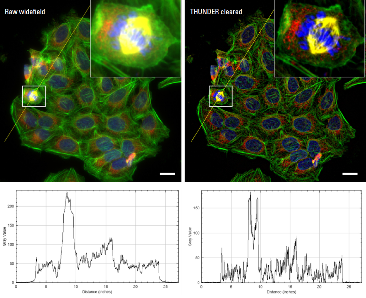
Figure 5: High resolution, high magnification images of U2OS human osteosarcoma cells, stained with DAPI (nuclei, blue), Alexa Fluor 488 Phalloidin (F‑actin, green), MitoTracker Red (mitochondria, red), and Alexa Fluor 647 Anti‑tubulin (tubulin, white). Acquired on THUNDER Imager Cell with 40x magnification. Left: Raw widefield image, Right: THUNDER cleared image. Scale bars are 50 µm. Both images show a magnified insert of the area highlighted by the white square. Below are line profiles across the yellow line through each image in the same location. U2OS cells provided by Dr. Niloufer Irani (Micron Bioimaging Facility, University of Oxford).
2D Imaging of HeLa Cell Cultures: Large FOV imaging
To further demonstrate the effects of THUNDER on the visualization of fine structural details, images were acquired from a different cell line (HeLa cells), labeled with different fluorescent labels. As shown in Figure 6, this sample shows smaller subcellular features, including detailed structures within each nucleus and the surrounding cytoskeleton, which were more clearly resolved following THUNDER computational clearing.
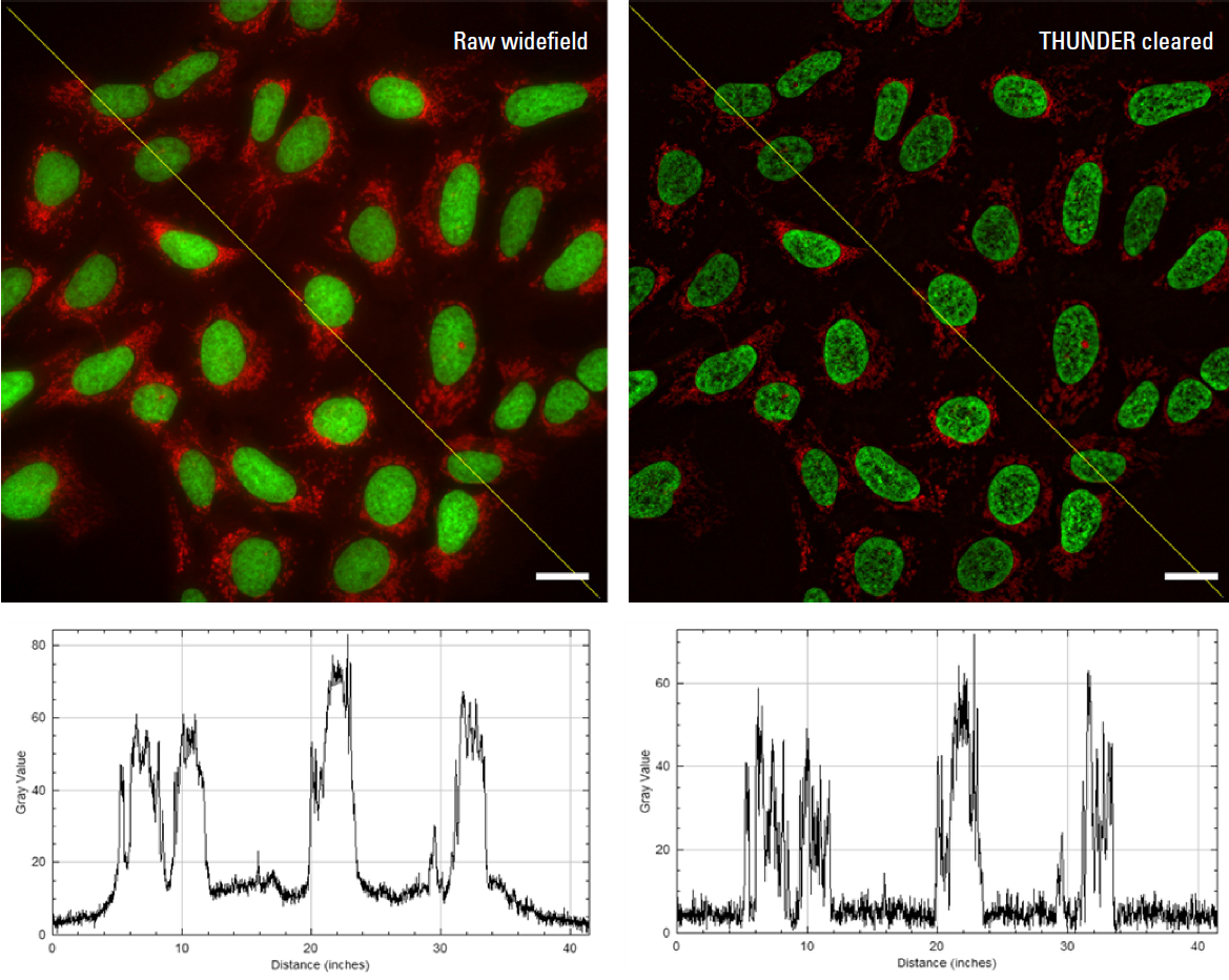
Figure 6: High resolution, high magnification images of U2OS human osteosarcoma cells, stained with DAPI (nuclei, blue), Alexa Fluor 488 Phalloidin (F‑actin, green), MitoTracker Red (mitochondria, red), and Alexa Fluor 647 Anti‑tubulin (tubulin, white). Acquired on THUNDER Imager Cell with 63x water immersion magnification. Left: Raw widefield image, Right: THUNDER cleared image. Scale bars are 20 µm. Below are line profiles across the yellow line through each image in the same location. HeLa cells provided by Dr. Niloufer Irani (Micron Bioimaging Facility, University of Oxford).
3D neural organoid imaging
In addition to fixed 2D cell cultures, THUNDER computational clearing offers significant benefits when working with large, complex 3D cultures such as organoids. To demonstrate this, a 1 mm diameter organoid composed of midbrain neuronal and non‑neuronal cells was imaged using the THUNDER Imager Cell with Kinetix22 (Figure 7). Owing to its thickness, this sample generated far more out‑of‑focus light than the 2D samples shown previously, presenting a more challenging sample for THUNDER to work on.
Figure 7 shows a representative 2D slice from the large 3D neural organoid, comparing the raw widefield image with the THUNDER computationally‑cleared image, alongside line profiles to show the quantitative effects on signal and background.

Figure 7: Large FOV 2D slices of a 1 mm diameter midbrain neural organoid stained with DAPI (blue, nuclear stain), β‑tubulin (green, neuronal stain), and GFAP (red, astrocyte stain). Acquired on THUNDER Imager Cell with 10x magnification. Left: Raw widefield image, Right: THUNDER cleared image. Scale bars are 400 µm. Below are line profiles across the yellow line through each image in the same location. Brain organoids provided by Dr. Tanya Singh (University of Oxford).
As thicker samples inherently generate more out‑of‑focus light than 2D cell samples, the effects of THUNDER computational clearing are more pronounced, resulting in substantial improvements in resolution and contrast. THUNDER was also applied to a 3D timelapse data set of the same neural organoid. A representative frame from this data set, showing the organoid viewed at an oblique angle relative to the imaging axis, is presented in Figure 8. This viewing angle further highlighted the impact of THUNDER on thick 3D samples, as it revealed structural features, positional information and morphology that was not discernable in the original raw widefield image.
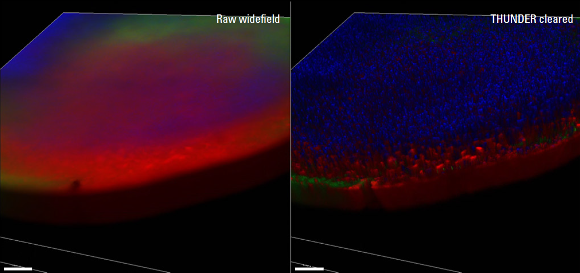
Figure 8: Large FOV 3D oblique view of a 1 mm diameter midbrain neural organoid stained with DAPI (nuclear stain), β‑tubulin (green, neuronal stain), and GFAP (red, astrocyte stain). Acquired on THUNDER Imager Cell with 10x magnification. Left: Raw widefield, Right: THUNDER cleared image. Scale bars are 400 µm. Brain organoids provided by Dr. Tanya Singh (University of Oxford).
Whole organism (Zebrafish) imaging
In the final example, a THUNDER Imager Cell with Kinetix22 was used to image zebrafish embryos (Figure 9). These large, complex samples are highly sensitive to light and require gentle illumination across a large field of view to capture the entire sample with good image quality. Figure 9 compares the raw widefield image with the THUNDER computationally‑cleared image, alongside line profiles to show the quantitative effects on signal and background.
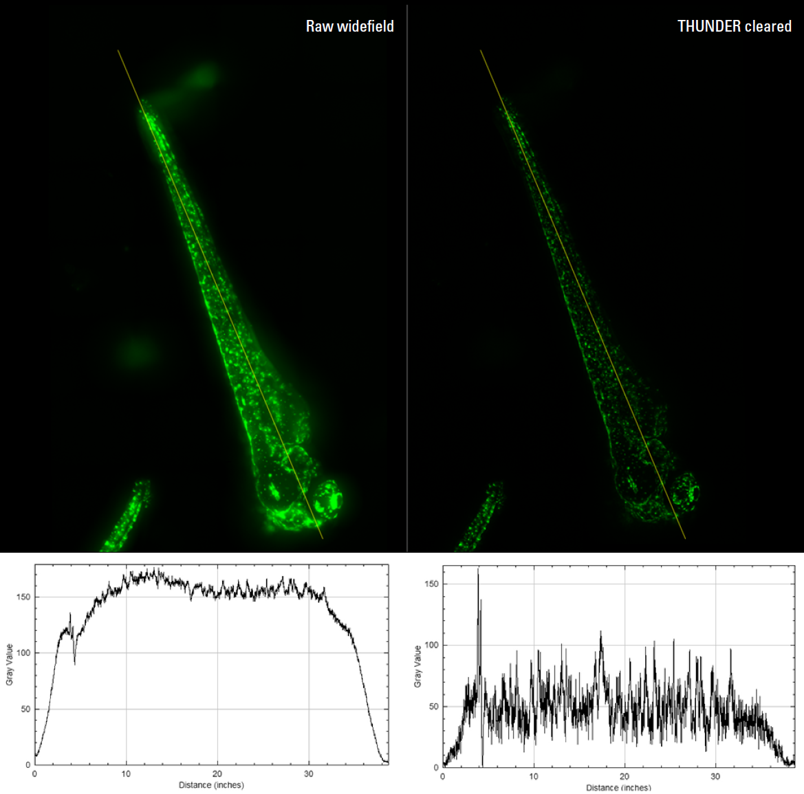
Figure 9: Large FOV image of an entire zebrafish embryo with GFP‑tagged melanocytes (green). Acquired on THUNDER Imager Cell with 10x magnification. Left: Raw widefield image, Right: THUNDER cleared image. Zebrafish embryos provide by the Richard White group (University of Oxford).
Discussion
The results demonstrate that the THUNDER Imager Cell system, combined with the Kinetix22 camera, can image across a large field of view while maintaining sub‑cellular resolution. The high sensitivity of the system enables reduced illumination levels and shorter exposure times to be used, helping to minimize photobleaching in fixed samples and phototoxicity in live‑cell experiments. Image quality and contrast are further enhanced using THUNDER computational clearing. By removing the blur and haze inherent to widefield images, this allows clear marking of signal and background for more accurate analysis. Together, this approach extends the applicability of widefield microscopy to optically challenging samples, complementing optical sectioning techniques.
Key benefits of combining THUNDER Imager Cell with Kinetix22:
> Matched FOV: Both the THUNDER Imager Cell and Kinetix22 feature a 22 mm field of view, optimizing image acquisition and enabling data capture from larger regions compared with typical CMOS cameras.
> Speed and sensitivity: The Kinetix22 sCMOS camera supports high-speed acquisition for imaging dynamic samples, while its low‑light sensitive imaging (due to 95% QE) and sub electron read noise levels, allow a reduction in both illumination intensity and exposure times.
> Resolution at magnification: As shown in this study, imaging was performed using a range of magnifications (10×, 20×, 40×, and 63× water immersion objectives). Across all magnifications, the THUNDER Imager Cell, combined with the Kinetix22 camera, delivered sub‑cellular resolution images with clear feature identification in both 2D and 3D samples.
> THUNDER Computational Clearing: This can be applied at any time during or after acquisition to generate high‑contrast, high‑resolution images across a wide range of challenging 2D and 3D samples.
> Intelligent automation: The THUNDER Imager Cell system integrates Adaptive Immersion for optimized water immersion objective performance, SmartCORR for correction of spherical aberrations at depth, and advanced triggering of the Kinetix22 sCMOS camera. Together with simplified experiment setup and navigation, it results in a powerful, easy‑to‑use automated imaging platform.
Conclusion
The THUNDER Imager Cell combined with the Kinetix22 sCMOS camera provides a robust, high‑speed platform for low‑dose, high‑contrast 2D and 3D imaging of large and complex biological samples. Together, these capabilities deliver a balanced, live cell friendly imaging solution well suited to discovery biology applications.
References
1. An Introduction to Computational Clearing https://www.leica‑microsystems.com/science‑lab/life‑science/an-introduction‑to‑computational‑clearing/
2. Leica Microsystems ‑ THUNDER Imager Cell ‑ Product Overview: https://www.leica‑microsystems.com/products/thunder‑imaging-systems/p/thunder‑imager‑cell/
3. Teledyne Photometrics Kinetix22 sCMOS Camera ‑ Product overview: https://www.teledynevisionsolutions.com/products/ kinetix/?model=01‑KINETIX‑22MM‑M‑C
4. Kinetix22 CMOS Camera Datasheet (rev. 2024): https://flir.netx.net/file/asset/60499/original/attachment
Materials and Methods
Samples
> U2OS cells were labelled with MitoTracker™ Red CMXRos (ThermoFisher Scientific, M7512; 250 nM) for 30 min at 37 °C, then fixed in 3% FA. Cells were processed for immunofluorescence (IF) using an anti tubulin primary antibody (DM1A) and Alexa FluorTM 647–conjugated anti mouse secondary antibodies. Nuclei were stained with DAPI (1 µg/mL), and F‑actin was labelled with Alexa Fluor 488 Phalloidin (165 nM, 10 min). Cells were washed in PBS and mounted in Mowiol DABCO.
> Fixed HeLa cells were labelled with GFP to mark nuclei and stained with MitoTracker Red CMXRos to label mitochondria.
> Human‑derived midbrain neural organoids (~1 mm in diameter) were labelled with DAPI to stain nuclei, β‑tubulin as a neuronal marker, and GFAP as an astrocytic marker. β‑tubulin and GFAP were detected using Alexa Fluor 488 and Alexa Fluor 594 conjugated antibodies.
> Live zebrafish larvae were labelled with GFP‑tagged melanocytes.
Imaging system
> Microscope: Leica Microsystems THUNDER Imager Cell system (a DMi8‑based platform) with SmartCORR and Adaptive Immersion technologies.
> Objectives: 10x objective (Ob. HC PL FLUOTAR 10x/0.32), 20x objective (HC PL APO 20x/0.80), 40x objective (HC PL APO 40x/0.95 CORR), 63x objective (Water Immersion 63x/1.2)
> Camera: Teledyne Photometrics Kinetix22 (22 mm diagonal sensor; 2400 × 2400 pixels; 6.5 µm pixel size; 95% QE; ~0.7 e‑ read noise; up to 500 fps full‑frame).
> Illumination: CoolLED pE‑800 LED light source with 8 channels and liquid light guide.
> Software: LAS X Life Science version 3.10
Acquisition settings
> Z‑stacks: 0.6‑1.0 µm steps; total depth 80–200 µm.
> Exposure: 10‑50 ms per channel.
> Illumination intensity: 10% or less.
> Dose minimization: combination of short exposure and low illumination, both leveraging the high sensitivity of the Kinetix22 camera.
> Images acquired in widefield configuration.
Image processing and analysis
THUNDER computationally‑cleared images were generated either during or after acquisition. Processed 3D datasets were analyzed using accurate deep‑learning based cell segmentation tools to extract morphological and quantitative parameters such as nuclei count, organoid volume, and lumen area (data not shown).
Acknowledgements
We would like to thank the following individuals for their support with this application note:
> Cell lines (U2OS and HeLa cells): Dr. Niloufer Irani, Micron Bioimaging Facility, University of Oxford.
> Brain organoids: Dr. Tanya Singh, Department of Physiology, Anatomy and Genetics, University of Oxford.
> Zebrafish embryos: Richard White group (Francisco López‑Cuevas and Vicky Tan), Oxford branch of the Ludwig Institute for Cancer Research, Nuffield Department of Medicine, University of Oxford.
> Data collection and workshop support: Deepthi Konthalapally and Abdullah Ahmed, Leica Microsystems.
> Imaging facilities and support: Niloufer Irani, Deirdre Kavanagh, and Lothar Schermelleh, Micron Bioimaging Facility, University of Oxford.
> Manuscript review: Dr. Sarah Piper, Dr. Jen Lee and Abdullah Ahmed.