3D Live Cell SCAPE
Prof. Alf Honigmann, Dr. Tom Borianne, Dr. Markus Mukenhirn, Pierre De Marinis
Center for Molecular and Cellular Bioengineering, TU Dresden, Germany
Background
The lab of Prof. Alf Honigmann focuses on the organisation and function of cell interfaces across scales. We spoke with postdoctoral researcher Dr. Markus Mukenhirn and Master’s student Pierre DeMarinis about their work in the Honigmann lab. Dr. Mukenhirn explained, “I work with mouse embryonic stem cells (mESCs) to study how they self-organise into 3D structures and the dynamics of their growth, observing them under the microscope over several days. By employing single-objective light sheet microscopy, we capture high resolution live-cell dynamics, revealing the intricate processes of lumen formation and tissue organization.”
The lab also explores other cell types, as Pierre describes, “I’ve been imaging MDCK kidney epithelial cells as they grow and form cysts, analysing how the lumen develops over time. These cells start as single cells and, within five days, self-organize into cysts about 80 microns in diameter, developing a central lumen — a hollow cavity within the 3D structure. In vivo, this process continues as the cyst elongates into tubular structures, eventually forming the renal system in the kidney.”
Pierre adds, “To minimize phototoxicity during long-term imaging of sensitive cells like stem cells and epithelial tissues, we use a SCAPE (swept confocally aligned planar excitation) microscopy setup. This allows us to gently illuminate the sample with low-power light while scanning at an angle in a light-sheet regime, enabling high-speed volumetric imaging without compromising cell viability.”
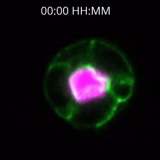
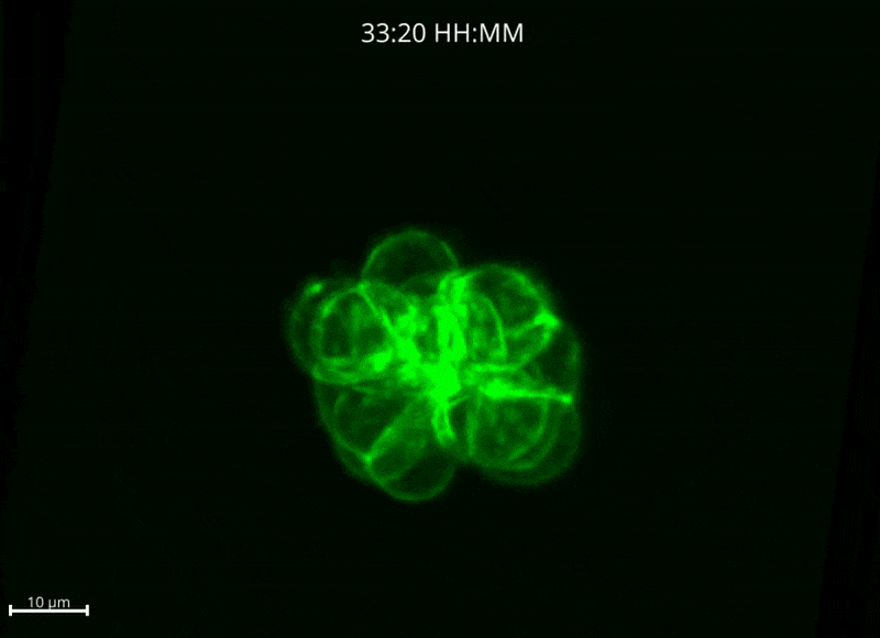
Figure 1: Top) 2D cross section time lapse video of MDCK cells. Bot) 3D time lapse video of mES cells. All images acquired by the Kinetix22 sCMOS camera.
Challenge
Dr. Mukenhirn outlined the imaging challenges they encounter in their research, “Our cells express three different fluorescent markers for live-cell imaging — cell membrane, lumen, and cell cell interface. One of the key challenges is imaging these markers simultaneously as the structures grow. We start with small formations that eventually expand up to 80 microns in diameter, so having a large camera sensor is a major advantage, allowing us to capture the entire structure throughout its development. We also want the flexibility to crop the central region of the sensor and mirror it later for specific analyses.”
He continued, "Currently, we capture a full 3D image stack every 10 minutes, which requires extremely low phototoxicity to keep the cells viable during long-term imaging. We use gentle illumination, but that results in a low signal, making high sensitivity essential. We're also planning calcium imaging assays, which will require capturing millisecond-scale stacks - so we'll need a high-speed camera to follow these rapid dynamics."
Pierre also mentioned, "We want to follow division events with our mouse embryonic stem cells (mES) and our MDCK cell line. We are using Napari and have developed our own algorithms in Python for image
analysis."
The Kinetix22 has significantly improved our light sheet live-cell imaging - its high sensitivity enables us to reduce phototoxicity and improve the quality and longevity of our imaging experiments, and push the temporal resolution when needed.
Dr. Markus Mukenhirn
Solution
The Kinetix22 is an ideal solution for this application, featuring ultra-high sensitivity across a large sensor, along with high speed and high dynamic range capabilities.
Dr. Mukenhirn described his experience with their two Kinetix22 cameras, “Since we are using two cameras, we have one for the green channel and one for the pink channel, we can image and mirror across both cameras simultaneously. We have two Kinetix22 light paths on our optical table with a beamsplitter, and we typically use Sensitivity mode, but we can switch to Dynamic Range mode when we have high and low signal levels in the same image, we like to have the added range.”
“The Kinetix22 is very good, you have very high sensitivity with helps us to be very gentle to our sample by lowering the illumination intensity, so we are able to image processes that couldn’t be imaged before because the phototoxicity was very high, so that’s been great.”
“We’ve also used this system to do DNA-PAINT, and the quantum yield is really excellent from these cameras.”