Imaging Fluorescent Microplastic Sedimentation
Prof. Stefan Peiffer and Marco La Capra
Hydrology Department, University Bayreuth, Bayreuth, Germany
Background
Prof. Dr. Stefan Peiffer's team at Bayreuth University’s Hydrology Department is exploring the interactions at the boundary between groundwater and surface water, which is vital for processes that influence the quality of both types of water. Prof. Peiffer’s research is focused on the efficiency and performance of matter processing, and the susceptibility of these systems to climate change and other human activity. The aim is to gain a deeper understanding of the interconnected physical and biogeochemical processes that govern the cycling of matter and energy at the interface between groundwater and surface water.
PhD student Marco La Capra's research focuses on microplastic transport in moving waters such as rivers. He described his work, “My main projects are analysing the sedimentation of microplastic geometries and the exfiltration of microplastics from sediments into water. In streambed sediments fluorescent microplastic particles of different states (beads, fibres, fragments) are imaged with fluorescence microscopy to determine the microplastic concentration.”
This research builds on recently published findings (Boos et al. 2024) and uses an experimental flume setup very similar to another publication (Boos et al. 2021).
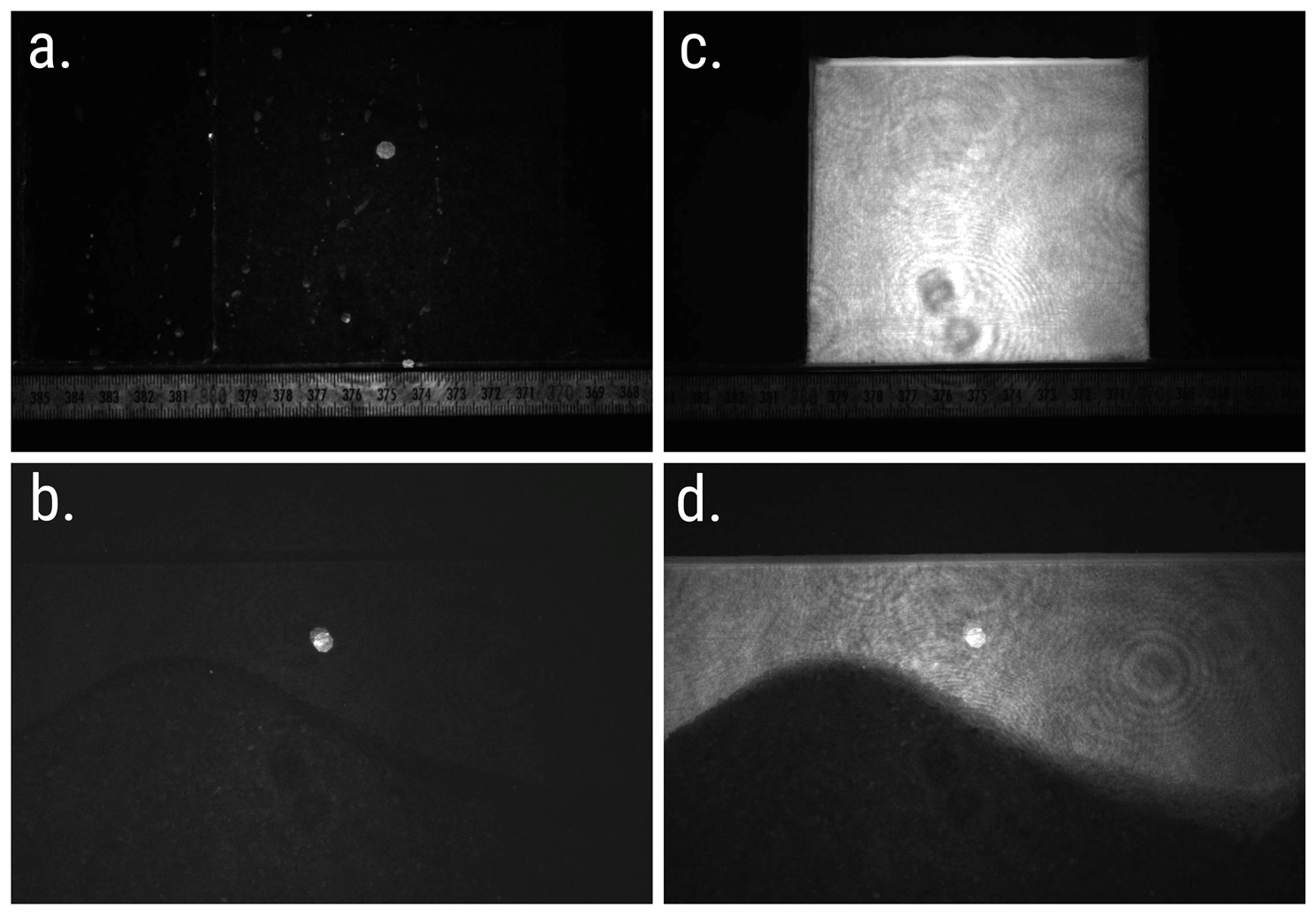
Figure 1: Calibration of the setup without (a) and with (b) a high concentration (2245 mg/L) of fluorescent microplastic in solution. c: Image of water before microplastic reaches the FOV. d: Image during the experiment. All images acquired with the Prime BSI Express sCMOS camera.
Challenge
Fluorescence imaging assays often require a high sensitivity scientific camera. The fluoresence imaging system used in Prof Peiffer's research is no exception, especially when the imaged microplastics are at a low concentration and challenging to detect. Furthermore, the quantitative nature of the experiment requires a reliable method to assign a quantity of fluorescent particles to a given grey value level. The observed processes also have to be imaged at different time scales, the used detector thus needs to be able to image fast and for longer periods of time.
The detector is required to be fast, sensitive, reliable and highly quantitative in order to make the best of this application.
We are more than happy with the quantum efficiency of the [Prime BSI Express], we also chose it because we knew the camera was perfect for imaging this kind of wavelength.
Marco La Capra
Solution
The Prime BSI Express is a well-balanced solution for this experimental setup. Marco explained, “The emission wavelength of the fluorescent microplastic particles is around 515 nm, this is close to the quantum efficiency maximum of the camera."
As the Prime BSI Express is back-illuminated it can image at ~500 nm with a near-perfect 95% quantum efficieny. Additionally, the readout noise of around one electron or less results in high signal to noise ratio images. The high sensitivity of the camera is paired with a linear behaviour between signal and gray values in the final image, which is paramount for quantitative studies, and the choice between sensitivity mode or high dynamic range (HDR) mode are optimal for the necessary acquisition speeds of around 20 fps and can still be increased up to 95 fps for more dynamic processes. With an array of 4.2 megapixels and a pixel size of 6.5 µm, the Prime BSI Express is very adaptable to various magnifications and enables users to reach optimal resolution in the experimental setup.
Learn More About The Prime BSI Express