In vivo Spinal Cord Calcium Imaging
Dr. Biafra Ahanonu
Department of Anatomy, University of California, San Francisco (UCSF), USA
Background
Dr. Biafra Ahanonu is a postdoctoral scholar and HHMI Hanna H. Gray Fellow in the lab of Prof. Alan Basbaum at UCSF, whose research primarily focuses on the neural and molecular mechanisms of pain. Dr. Ahanonu describes his work, “We are trying to understand how the nervous system changes in response to acute and chronic pain. If we can identify which neurons change, we can potentially modulate those neurons to reduce pain.”
“We study the brain and spinal cord and are using fluorescent calcium imaging to infer the state of different neuronal and glial populations in awake, behaving animal models. We observe spontaneous neural activity during baseline imaging and then we study evoked neural activity by stimulating with von Frey (stiff bristle) hairs or other stimuli, like those that produce hot or cold sensations. Further, while a stimulus can be the same, the perception of that stimulus can change over time or in chronic pain. We would like to answer: where in the nervous system is that change happening?”
Figure 1: Spinal cord calcium imaging in an awake, behaving animal during delivery of peripheral stimuli. Spinal cord projection neuron activity (Phox2a-Cre; Ai162 (GCaMP6s)) aligned to stimulus application (red vertical lines at bottom). Field of view is 2169 x 3036 µm. Images acquired by Kinetix sCMOS camera.
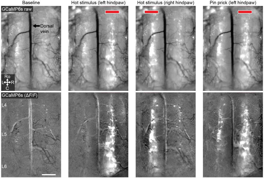
Figure 2: Spinal cord calcium imaging in an awake, behaving animal during delivery of peripheral stimuli. Spinal cord projection neuron activity (Phox2a-Cre; Ai162 (GCaMP6s)) aligned to stimulus application on a single side of the body (red bars). Scale bar, 300 µm; field of view is 2169 x 3036 µm. Images acquired by Kinetix sCMOS camera.
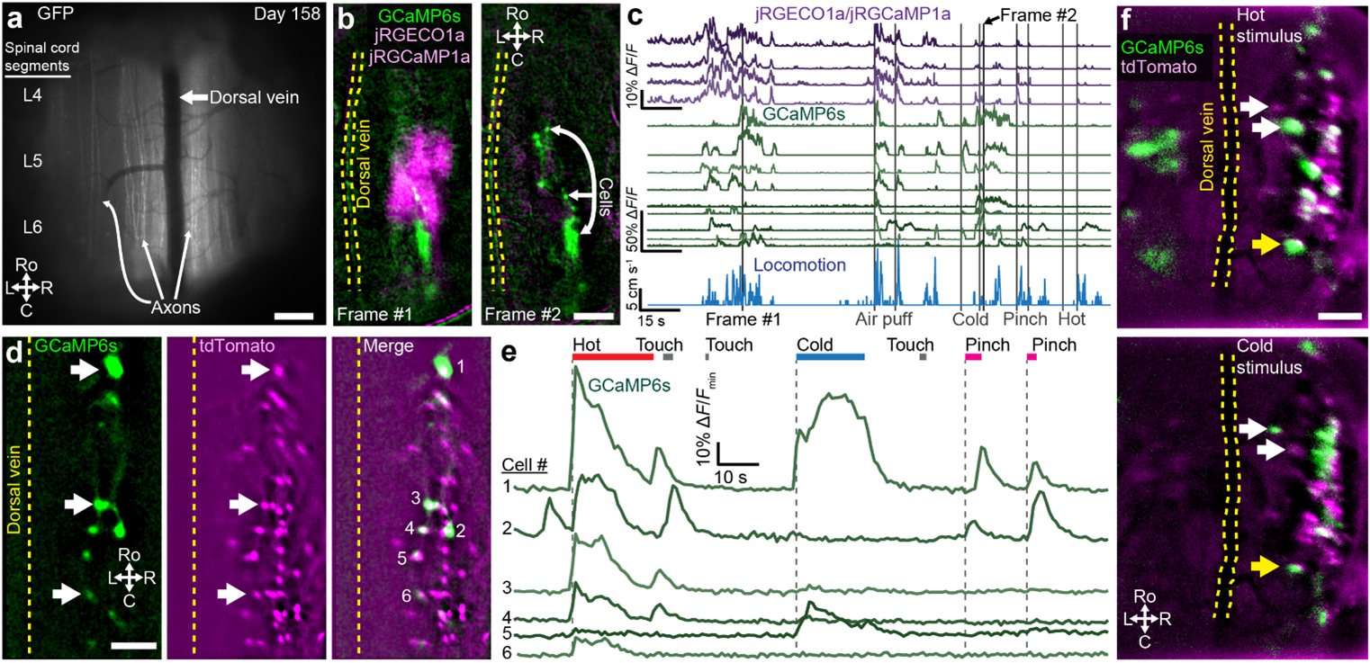
Figure 3: Spinal cord imaging of axons and neural activity with the Kinetix sCMOS camera. A) Image of multiple segments of the left and right spinal cord showing GFP+ axons on the 158th day of imaging a Thy1-GFP mouse. B) Simultaneous imaging using a TwinCam and two Kinetix cameras as animal locomotes (Frame #1) or responds to a cold stimulus (Frame #2) of red (jRGECO1a and jRGCaMP1a) and green (GCaMP6s) Ca2+ indicators. C) Quantification of calcium transients from multiple regions (magenta) or cells (green) as animal responds to sensory stimuli (vertical lines) or locomoted (blue trace, running wheel attached to a rotary encoder). D) Simultaneous imaging of spinal cord projection neuron (SCPN) activity (green) and an always-visible marker of SCPNs (magenta) in a Phox2a-Cre; Ai162 (GCaMP6s, green); Ai9 (tdTomato, magenta) mouse. White arrows, active tdTomato+ cells. E) Quantification of calcium transients from cells numbered as in D in response to various peripheral stimuli applied to the hind paw. F) Same as D in another animal. Scale bars: 200 μm. The anatomy axes abbreviations: C, caudal; L, left; R, right; Ro, rostral. Neural activity extracted with CIAtah (github.com/bahanonu/ciatah) using CELLMax (https://doi.org/10.25740/vh359hb5216) or ROI analysis.
Challenge
Dr. Ahanonu explained his imaging challenges, “We need rapid, sensitive acquisition for multiple reasons, such as imaging fast calcium transients, reducing phototoxicity and bleaching, and handling the spinal cord’s unique motion compared to most brain imaging. During imaging, the spinal cord can move hundreds of microns very rapidly, in under a second, necessitating very short exposure times to avoid a blurry image and for easier motion correction during analysis. Thus, while we may acquire images at 20 hertz, we need an exposure time between 1–10 milliseconds. As these low exposure times make it harder to detect weaker, biologically relevant signals, the read noise and quantum efficiency then becomes critical. The higher read noise and lower quantum efficiency of our older cameras made it hard to detect signals without averaging or other compromises, such photodamage caused by using higher excitation power when trying to increase SNR, so we needed a more sensitive camera.”
“There are times when an injury occurs on just one side of the body and pain perception spreads to other parts of the body. Thus, we want a large field of view to capture both sides of the spinal cord. We can then address: if we give a stimulus on the left side of the body how does it affect both sides of the spinal cord? A larger field of view also helps us image multiple segments of the spinal cord that are activated by stimulating different body parts, so it is a key feature.”
Dr. Ahanonu requires a detector that features high speed acquisition, high sensitivity with low read noise, and all paired with a large field of view..
The Kinetix has been instrumental in imaging at high SNR to detect even weaker cells, and the multiple imaging modes are really useful.
Dr. Biafra Ahanonu
Solution
The Kinetix sCMOS is the ideal imaging solution for this application, and Dr. Ahanonu uses multiple for this project, “We tested multiple different cameras from several companies on the same systems to get a fair comparison and chose the Kinetix. The Kinetix has been great overall, in terms of the large field of view and having multiple imaging modes, which has been really useful. I have primarily used either Sensitivity or Dynamic Range modes depending on the intensity of the fluorescent indicator. And I have also used Speed mode in certain contexts, for example, when looking at vasculature and wanting to acquire at 500 to 900 Hz.”
“It has been a positive experience; we often use the Kinetix system because of the added advantages of the high quantum efficiency and the low read noise. The Kinetix has been instrumental in this project, allowing us to get the exposure rate low enough for the motion correction while still obtaining a high enough signal to detect low-SNR cells.”
“The two Kinetix cameras are integrated into a 3i VIVO multiphoton microscope using a TwinCam. We use one Kinetix for our red channel and one for green so we can do simultaneous multicolour imaging across the large field of view. This is running in 3i’s SlideBook imaging software, but we also use Photometrics software for simultaneous recordings. We send TTL triggers to sync the Kinetix cameras.”
“Voltage imaging is something that we are interested in, we can use the Kinetix in combination with holographic excitation to do fast, high-SNR voltage imaging potentially in the future.”
Reference
Ahanonu*, Crowther*, et al. (2024) Long-term optical imaging of the spinal cord in awake behaving mice. Nature Methods. https://doi.org/10.1038/s41592-024-02476-3.