Large-scale Antimicrobial Single-Cell Testing
Dr. Lucas Boeck
Department of Biomedicine, University of Basel, Basel, Switzerland
Background
Microbial drug tolerance is a major threat to global health, referring to the ability of bacteria to survive antibiotic exposure without any genetic resistance mechanisms, regrowing once the drug is cleared. In mycobacterial infections, this challenge is particularly acute, Mycobacterium tuberculosis claimed more than one million lives in 2023 alone and treatment regimens still require a minimum of four months.
Standard antibiotic susceptibility and drug tolerance testing relies on measuring minimum inhibitory concentrations (MICs), namely the drug concentration that prevents bacterial growth, but growth inhibition does not capture the killing dynamics that ultimately determine whether an infection is cleared. Colony-forming unit (c.f.u.) assays can measure killing, but they are labour-intensive, low-throughput, and provide only a snapshot in time.
To address this fundamental gap, Dr. Lucas Boeck, research group leader at the Department of BIomedicine at the University of Basel Switzerland, developed antimicrobial single-cell testing (ASCT), a highly scalable live-cell imaging platform that quantifies bacterial viability in real time, at single-cell resolution, across thousands of conditions simultaneously. The platform was applied first to M. tuberculosis to evaluate whether killing dynamics predict drug regimen efficacy in mice and humans, and then extended to 405 clinical M. abscessus isolates to determine whether strain-specific killing behaviour is associated with individual patient outcomes.
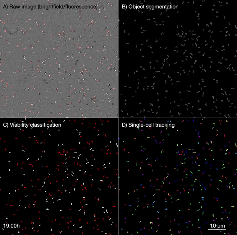
Figure 1: Automated image analysis in Antimicrobial Single-Cell Testing (ASCT). Raw brightfield images (cell morphology; 100x objective) and fluorescence images (quantifying propidium iodide [PI] accumulation) are processed to segment cells (lines indicate cell borders), classify bacterial viability (white: viable; red: dead) and track individual cells (track indicated by colours) over 72 hours. Image is a still frame taken from source video from Jopanovic et al 2026: https://static-content.springer.com/esm/art%3A10.1038%2Fs41564-025-02217-y/MediaObjects/41564_2025_2217_MOESM4_ESM.mp4
Challenge
The central imaging challenge of ASCT is one of extraordinary scale combined with demanding sensitivity requirements. Each experiment tracks the viability of millions of individual bacteria across up to 1,536 wells simultaneously, capturing brightfield and fluorescence images of over 10,000 fields every 2-4 hours for up to 7 days, generating up to one million images per experiment with datasets reaching 11 TB in size. At the heart of the viability assay is propidium iodide (PI), a cell-impermeable fluorescent dye that accumulates in bacteria only upon membrane disruption, serving as a proxy for cell death. Detecting PI fluorescence reliably at the single-cell level in immobilised mycobacteria, rod-shaped cells just 1–3 µm in size, demands a camera with exceptional sensitivity, speed, and signal-to-noise performance.
Critically, the platform must maintain consistent fluorescence quantification across all well positions and all timepoints throughout multi-day experiments, any spatial or temporal variation in background fluorescence would confound the single-cell PI classifications that underpin the entire analysis. The platform must also operate at sufficient throughput to image 9 fields per well across up to 1,536 wells within ~130 minutes per timepoint, this demands rapid, high-fidelity image acquisition without sacrificing the sensitivity needed to reliably classify individual live and dead bacteria.
By studying millions of single-cell fates across hundreds of conditions, our approach provides a scalable framework to translate in vitro killing into in vivo efficacy, opening new avenues for drug development and personalized therapy.
Dr. Lucas Boeck
Solution
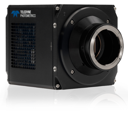
The Kinetix sCMOS camera is the ideal solution for this application, Dr. Boeck and team made use of the Kinetix’s high quantum efficiency, low read noise, and rapid imaging speed across a range of magnifications. The Kinetix met the two main demands of ASCT, capturing faint PI fluorescence signal from individual microscale bacteria, while sustaining the throughput required to image millions of cells across a 1,536-well plate within a strict imaging window.
The performance of the Kinetix enabled high automated classification accuracy (AUC-ROC = 0.9989) when validated against manually annotated ground-truth datasets. This level of accuracy was essential for building the ~20,000 time–kill curves that form the analytical backbone of the study. Kinetix performance across the full 72 hour M. abscessus and 168 hour M. tuberculosis imaging runs ensured that data quality remained consistent from the first to the final timepoint, resulting in the robust quantification of drug tolerance phenotypes across 405 clinical isolates. In total, the ASCT platform enabled the tracking of over 140 million individual mycobacteria, a feat only achievable with a detector capable of delivering both the sensitivity and the sustained throughput that the Kinetix provides.
Reference
Jovanovic, A., Bright, F.K., Sadeghi, A., Wicki, B., Caño Muñiz, S.E., Giannini, G.C., Toprak, S., Sauteur, L., Rodoni, A., Wüst, A., Lupien, A., Borrell, S., Grogono, D.M., Wheeler, N.E., Dehio, P., Nemeth, J., Pargger, H., Thomson, R., Bell, S.C., Gagneux, S., Bryant, J.M., Peng, T., Diacon, A.H., Floto, R.A., Abanto, M. & Boeck, L. (2026). Large-scale testing of antimicrobial lethality at single-cell resolution predicts mycobacterial infection outcomes. Nature Microbiology, 11, 566–583. https://doi.org/10.1038/s41564-025-02217-y